D. Renovaldi, M.Sc.
Editorial Office, The ACTO Times
International PhD Program in Medicine, Taipei Medical University, Taipei, Taiwan
Abstract
Neutrophils are traditionally portrayed as short-lived innate immune cells that rapidly migrate to sites of infection or tissue injury. Cancer, however, can appropriate these emergency-response programs for its own benefit. Tumor-derived signals alter granulopoiesis, neutrophil maturation, trafficking, lifespan, and activation, generating heterogeneous neutrophil populations with markedly different functions. Some neutrophils retain antitumor activity through direct cytotoxicity, antibody-dependent killing, and support of adaptive immune responses. Others promote tumor growth by stimulating angiogenesis, remodeling the extracellular matrix, suppressing T-cell activity, escorting circulating tumor cells, establishing premetastatic niches, and releasing neutrophil extracellular traps. The historical N1–N2 polarization model provided an important framework for understanding this functional plasticity, but single-cell and spatial studies now reveal a continuum of maturation and activation states shaped by tumor type, anatomical location, disease stage, and treatment. Importantly, anticancer therapy may either reinforce suppressive neutrophil programs or generate interferon-responsive, tumor-controlling populations. This review traces the changing perception of neutrophils in cancer and discusses why selectively targeting harmful neutrophil programs may be more effective than indiscriminate neutrophil depletion.
Keywords: neutrophils; tumor microenvironment; tumor-associated neutrophils; neutrophil extracellular traps; metastasis; immunotherapy
The original script: arrive, attack, and repair
Neutrophils are the most abundant circulating leukocytes in humans and constitute one of the earliest cellular responses to infection and tissue damage. Guided by chemotactic signals, they rapidly exit the circulation and deploy antimicrobial mechanisms that include phagocytosis, reactive oxygen species, proteolytic granules, and neutrophil extracellular traps (NETs) [1]. These responses are powerful but potentially destructive; neutrophils must therefore be rapidly recruited, tightly regulated, and efficiently removed after completing their task.
Under physiological conditions, neutrophils contribute to immune surveillance through direct cytotoxicity, reactive oxygen species production, degranulation, and interactions with adaptive immune cells. In cancer, however, these protective functions may be progressively redirected toward tumor-supportive activities (Figure 1).
Cancer disrupts this normally transient response. A growing tumor resembles a wound that does not resolve, continuously producing inflammatory mediators, damage-associated signals, hypoxia, and necrotic material. These stimuli sustain neutrophil production and recruitment while exposing the cells to a microenvironment capable of changing their phenotype. Instead of merely entering tumors as terminally differentiated bystanders, neutrophils may be reprogrammed during development in the bone marrow, while circulating in the blood, and again after entering tumor tissues [2–6]. The major stages of this transition from homeostatic first responders to tumor-educated accomplices are summarized in Figure 1.
This recognition fundamentally changed the view of neutrophils in oncology. Tumor-associated neutrophils (TANs) are now understood as active components of the tumor ecosystem.
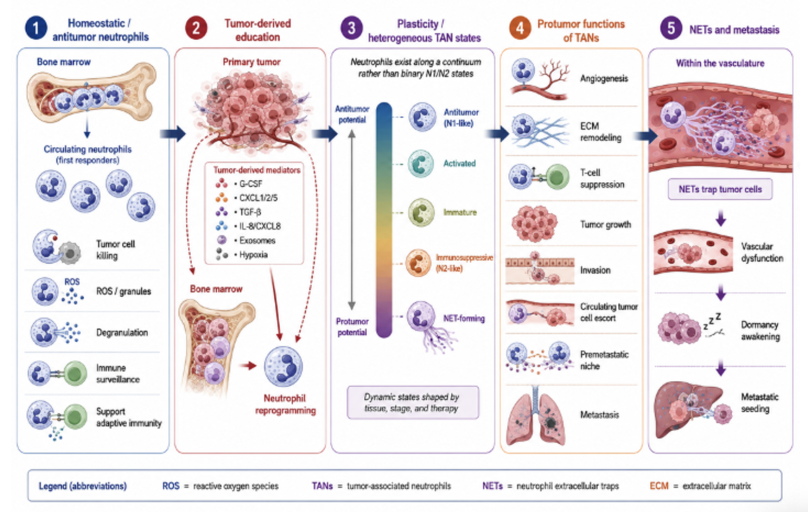
Figure 1. Neutrophils in cancer progression: from host defense to tumor support
Their contribution is not uniformly beneficial or harmful. Depending on context, neutrophils can destroy malignant cells, promote immune surveillance, or become potent facilitators of tumor progression (Figure 1).
Beyond N1 and N2: neutrophils as changing cellular states
An influential early model divided TANs into antitumor “N1” and protumor “N2” phenotypes. In murine tumors, transforming growth factor-β promoted an N2-like state, whereas its inhibition generated hypersegmented, inflammatory neutrophils with enhanced tumor-killing properties [2]. The model was conceptually valuable because it established that neutrophil behavior was plastic rather than predetermined.
Subsequent studies nevertheless showed that neutrophil biology cannot be fully represented by two stable categories. Differences in maturation, density, tissue location, metabolism, circadian age, and exposure to cytokines all influence neutrophil function [3–6]. Neutrophil phenotypes are therefore better understood as a functional continuum extending from cytotoxic and immunostimulatory states to immature, suppressive, NET-forming, and prometastatic states (Figure 2).
Low-density neutrophils, for example, include both immature immunosuppressive cells and activated mature cells, demonstrating that physical separation or a limited marker panel cannot reliably define biological function [3]. Considerable overlap also exists between immunosuppressive neutrophils and polymorphonuclear myeloid-derived suppressor cells.
In many settings, these terms describe intersecting functional states rather than completely distinct cellular lineages [4–6].
Neutrophil heterogeneity can arise at several points along their developmental trajectory. Tumor-derived cytokines and chemokines may alter granulopoiesis in the bone marrow, promote the premature release of immature neutrophils, and further modify mature neutrophils in the circulation or tumor microenvironment. Hypoxia, transforming growth factor-β, interferons, metabolic stress, and anticancer therapy provide additional layers of regulation (Figure 2).
Single-cell RNA sequencing has provided a more detailed map of this heterogeneity. Analyses across human and murine lung cancers identified conserved neutrophil populations as well as species- and tissue-specific states [7]. In hepatocellular carcinoma, single-cell analysis revealed distinct trajectories from immature to mature neutrophils, including populations associated with poor prognosis and tumor-promoting interactions [8]. These findings suggest that neutrophils should be interpreted as a dynamic spectrum generated by developmental history and local instruction (Figure 2).
Spatial context is equally important. A neutrophil positioned in the circulation, at the invasive margin, within a hypoxic tumor core, or near metastatic vessels encounters different signals and cellular partners. Consequently, the same marker may not indicate the same function across compartments.
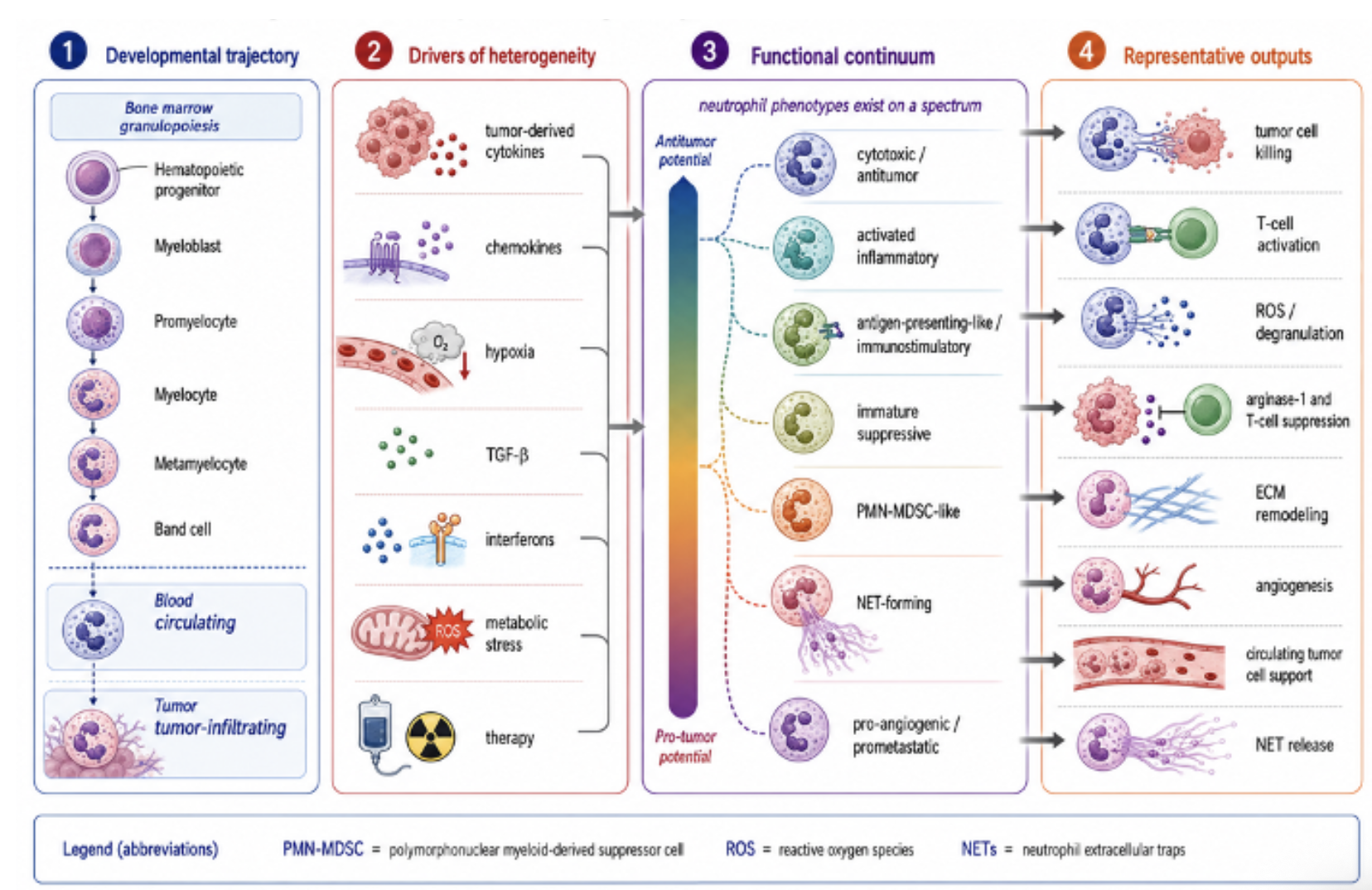
Figure 2. Neutrophils heterogeneity and functional states in cancer
This complexity helps explain why high neutrophil abundance or an elevated blood neutrophil-to-lymphocyte ratio is frequently associated with unfavorable outcomes but cannot, by itself, establish what the neutrophils are doing [9].
How tumors recruit and corrupt first responders
Tumors influence neutrophils before the cells reach the primary lesion. Cancer-derived granulocyte colony-stimulating factor, CXCL-family chemokines, inflammatory cytokines, metabolites, and extracellular vesicles can stimulate emergency granulopoiesis and release immature neutrophils into the circulation [4–6]. Once recruited through pathways such as CXCL1/2/5–CXCR2, neutrophils are exposed to hypoxia, transforming growth factor-β, interleukins, lipid mediators, and tumor-derived extracellular vesicles. These interconnected mechanisms of neutrophil recruitment and reprogramming are illustrated in Figure 1.
Nevertheless, antitumor neutrophil activity remains biologically possible. In experimental models, neutrophils recruited to the lung before metastatic colonization directly eliminated disseminated tumor cells through hydrogen peroxide-dependent mechanisms [10]. Neutrophils can also inhibit tumor growth by releasing the receptor tyrosine kinase MET in response to tumor-derived signals, illustrating how pathways commonly associated with cancer cells may produce opposite effects in immune cells [11]. Additional antitumor mechanisms include antibody-dependent cellular cytotoxicity, production of inflammatory cytokines, recruitment of lymphocytes, and interactions that support T-cell activation (Figures 1 and 2).
The balance often shifts as tumors progress. Tumor-conditioned neutrophils release vascular endothelial growth factor, matrix metalloproteinases, elastase, and other granule components that promote angiogenesis, extracellular-matrix degradation, invasion, and growth-factor availability. They may suppress T-cell responses through arginase-1, reactive oxygen species, nitric oxide-related pathways, checkpoint ligands, and competition for essential metabolites [4–6]. The principal protumor outputs of TANs, including angiogenesis, extracellular-matrix remodeling, T-cell suppression, tumor-cell invasion, and metastatic support, are summarized in Figures 1 and 2.
In breast cancer models, interleukin-17-producing γδ T cells induced systemic neutrophil expansion, and these neutrophils suppressed cytotoxic CD8-positive T cells, thereby facilitating metastasis [12]. Cancer therefore does not necessarily create a completely new neutrophil program. Rather, it redirects normal emergency functions. Proteases intended to clear damaged tissue can open invasion paths. Oxidants designed to kill microorganisms can suppress lymphocytes or damage surrounding tissue. Proangiogenic and repair signals intended to restore perfusion can feed a growing tumor. Mechanisms that are protective during acute injury become harmful when activated continuously (Figure 1).
Accomplices in the metastatic event
Neutrophils can assist cancer cells at nearly every stage of metastasis. At the primary tumor, they promote invasion and vascular entry. In the circulation, they form heterotypic clusters with circulating tumor cells.
Analysis of patients with breast cancer showed that neutrophil-associated circulating tumor cells had enhanced proliferative activity and a greater capacity to generate metastases than isolated tumor cells [13]. Thus, neutrophils may operate not only as passive shields but also as cellular escorts that provide survival and growth signals (Figure 1).
Distant organs may also be conditioned before tumor cells arrive. Tumor-derived signals recruit neutrophils to future metastatic sites, where they remodel extracellular matrix, alter vascular permeability, and create inflammatory niches. In breast cancer models, neutrophils accumulated in the premetastatic lung and promoted colonization through leukotriene production, selectively supporting highly tumorigenic cancer-cell subclones [14].
NET formation is one of the clearest examples of an antimicrobial mechanism being repurposed by cancer. NETs consist of decondensed chromatin decorated with histones, elastase, myeloperoxidase, and other granule proteins [15]. They were originally characterized as structures that immobilize microorganisms, but cancer-associated NETs can capture circulating tumor cells, facilitate adhesion to vascular or tissue surfaces, and promote metastatic seeding [16]. NET-associated proteases can also remodel extracellular matrix proteins and awaken dormant cancer cells, providing a potential link between inflammation and late metastatic relapse [17].
NETs additionally connect cancer with thrombosis and vascular dysfunction. Tumor-bearing mice develop systemic neutrophil priming and increased NET formation, which can contribute to cancer-associated hypercoagulability [15]. More recently, neutrophils and NETs were shown to obstruct tumor-associated blood vessels, generating necrosis while paradoxically increasing metastatic dissemination [23]. These findings reveal a counterintuitive process: neutrophil-mediated vascular damage may destroy parts of a tumor yet simultaneously select for aggressive cells and create routes for escape. The proposed sequence linking NET-mediated tumor-cell trapping, vascular dysfunction, dormancy awakening, and metastatic seeding is shown in Figure 1.
NETs additionally connect cancer with thrombosis and vascular dysfunction. Tumor-bearing mice develop systemic neutrophil priming and increased NET formation, which can contribute to cancer-associated hypercoagulability [15]. More recently, neutrophils and NETs were shown to obstruct tumor-associated blood vessels, generating necrosis while paradoxically increasing metastatic dissemination [23].
These findings reveal a counterintuitive process: neutrophil-mediated vascular damage may destroy parts of a tumor yet simultaneously select for aggressive cells and create routes for escape.
Treatment as a second rewiring event
Anticancer treatment does not merely eliminate tumor cells; it changes the inflammatory environment in which neutrophils develop and function. Chemotherapy-induced cell death can release damage-associated molecules and cytokines that recruit or activate neutrophils. In some tumors, this response contributes to resistance. Chemotherapy was shown to induce NET formation and activate latent transforming growth factor-β, creating an immunosuppressive environment that reduced treatment efficacy [19].
The relationship is not universal. In a colorectal cancer model, chemotherapy-induced NETs inhibited tumor growth rather than promoting it [20]. Although apparently contradictory, these observations reinforce the central principle that neutrophil functions are context-dependent. Tumor origin, microbial exposure, treatment type, NET composition, timing, and the surrounding immune landscape may determine whether the resulting response supports tumor control or recovery.
Immunotherapy provides further evidence that neutrophils can be therapeutically redirected. In preclinical cancer models, successful immunotherapy induced an interferon-stimulated neutrophil state with enhanced antitumor activity [21]. A related study identified IRF1-dependent neutrophils that were required for effective immunotherapy and associated with favorable responses in patients [22]. These observations support therapeutic strategies that reprogram neutrophils toward antitumor states rather than eliminating the entire neutrophil population (Figure 3).
Neutrophils may consequently serve as both mediators and biomarkers of treatment response. A high circulating neutrophil count is often interpreted as evidence of suppression, yet numerical abundance does not reveal cellular state. Interferon-responsive mature neutrophils, immature suppressive neutrophils, senescent cells, and NET-forming neutrophils could have opposite effects despite contributing to the same total count. Biomarker strategies will therefore need to combine abundance with maturation, transcriptional state, activation, tissue localization, and functional assays.
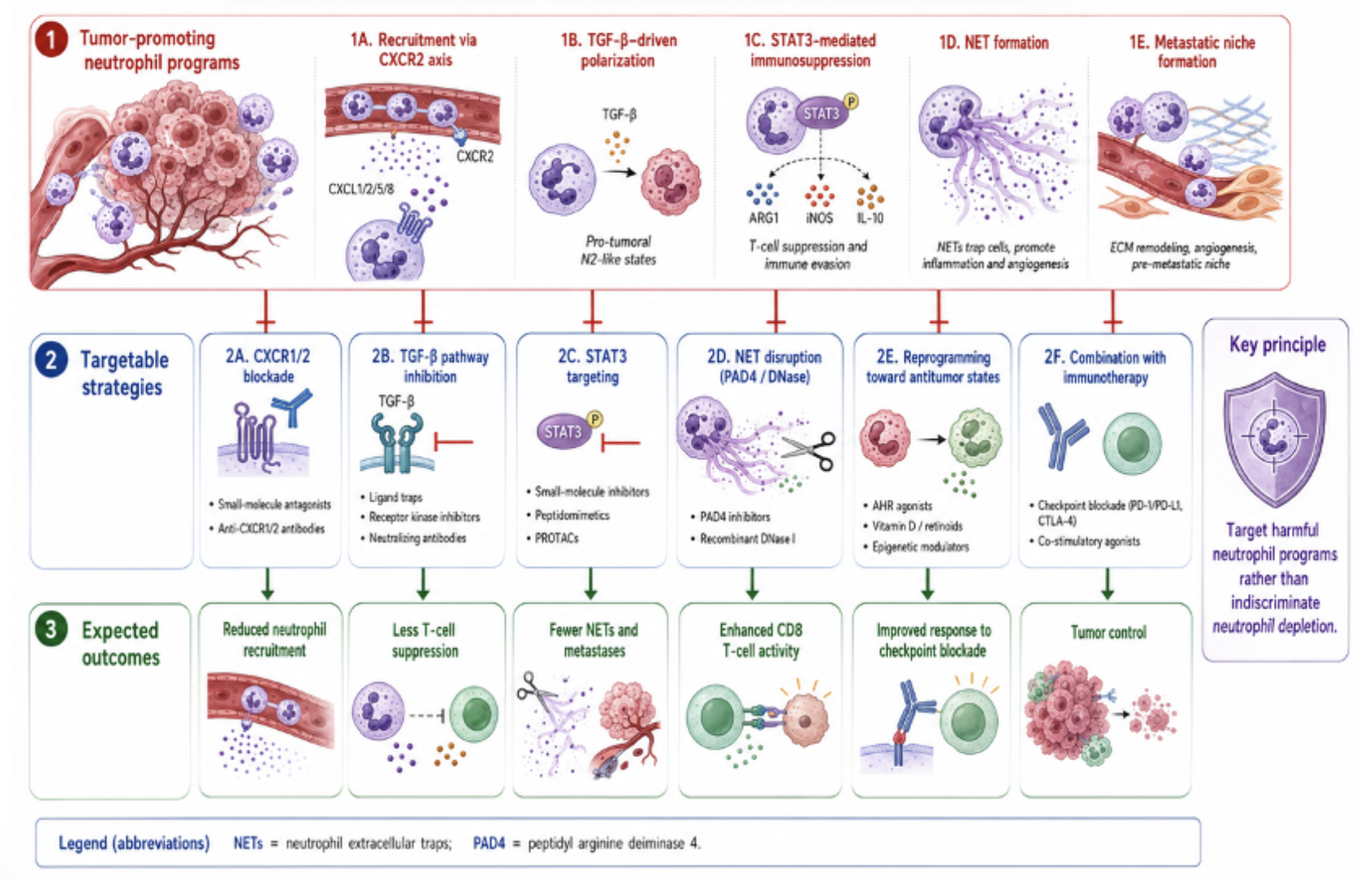
Figure 3. Therapeutic targeting of neutrophil programs in cancer
Target the program as the main strategy
The therapeutic appeal of neutrophils is accompanied by an obvious risk. Global neutrophil depletion would compromise antimicrobial defense and tissue repair. A more rational objective is to interrupt the specific pathways through which tumors recruit, educate, or exploit neutrophils.
Potential approaches include CXCR1/2 blockade to restrict tumor-directed neutrophil recruitment, inhibition of transforming growth factor-β or STAT3-dependent suppressive programs, disruption of NET formation using PAD4 inhibitors or DNase-based strategies, and reprogramming of neutrophils toward antitumor phenotypes [5,6,18]. These strategies and their expected effects on T-cell activity, NET formation, metastasis, and checkpoint immunotherapy are summarized in Figure 3.
Recent neutrophil-specific STAT3 targeting reduced tumor progression and expanded cytotoxic CD8-positive T-cell responses, supporting the feasibility of selectively disabling suppressive neutrophil programs [24]. Other strategies may seek to enhance Fc receptor-mediated cytotoxicity or preserve interferon-responsive neutrophils during immunotherapy. Combining neutrophil-directed interventions with immune-checkpoint blockade may therefore improve antitumor immunity while avoiding the infectious risks associated with indiscriminate neutrophil depletion (Figure 3). Translation will require more precise definitions. Many mechanistic studies rely on mouse models, whereas human neutrophils differ in marker expression, lifespan, and sensitivity to experimental manipulation.
Sample processing can itself activate neutrophils or induce changes in density, transcription, and NET formation. Prospective human studies should therefore integrate blood and tissue analysis, spatial imaging, single-cell or proteomic profiling, and longitudinal sampling before and after treatment [5,6,25].
Conclusion
The story of neutrophils in cancer has evolved from one of simple recruitment to one of profound cellular adaptation. These cells are neither inherently tumor-promoting nor reliably antitumor. They are rapid-response effectors whose normal functions killing, proteolysis, vascular modification, immune regulation, and tissue repair can be redirected by malignant tissues.
The N1–N2 framework first captured this plasticity, but current evidence supports a multidimensional continuum shaped by maturation, anatomy, tumor stage, systemic inflammation, and therapy. Neutrophils may kill disseminated cells in one setting, suppress lymphocytes in another, and change function again following chemotherapy or immunotherapy. NETs similarly illustrate how the same biological mechanism can promote thrombosis, dormancy escape, treatment resistance, or, under selected conditions, tumor control.
The central therapeutic challenge is therefore not to remove neutrophils indiscriminately. It is to identify when, where, and how they become tumor accomplices and to selectively disarm those programs while preserving or restoring their capacity to defend the host.
References
- Rosales C. Neutrophil: a cell with many roles in inflammation or several cell types? Front Physiol. 2018;9:113.
- Fridlender ZG, Sun J, Kim S, Kapoor V, Cheng G, Ling L, et al. Polarization of tumor-associated neutrophil phenotype by TGF-β: “N1” versus “N2” TAN. Cancer Cell. 2009;16(3):183–194.
- Sagiv JY, Michaeli J, Assi S, Mishalian I, Kisos H, Levy L, et al. Phenotypic diversity and plasticity in circulating neutrophil subpopulations in cancer. Cell Rep. 2015;10(4):562–573.
- Jaillon S, Ponzetta A, Di Mitri D, Santoni A, Bonecchi R, Mantovani A. Neutrophil diversity and plasticity in tumour progression and therapy. Nat Rev Cancer. 2020;20(9):485–503.
- Hedrick CC, Malanchi I. Neutrophils in cancer: heterogeneous and multifaceted. Nat Rev Immunol. 2022;22(3):173–187.
- Shaul ME, Fridlender ZG. Tumour-associated neutrophils in patients with cancer. Nat Rev Clin Oncol. 2019;16(10):601–620.
- Zilionis R, Engblom C, Pfirschke C, Savova V, Zemmour D, Saatcioglu HD, et al. Single-cell transcriptomics of human and mouse lung cancers reveals conserved myeloid populations across individuals and species. Immunity. 2019;50(5):1317–1334.e10.
- Xue R, Zhang Q, Cao Q, Kong R, Xiang X, Liu H, et al. Liver tumour immune microenvironment subtypes and neutrophil heterogeneity. Nature. 2022;612(7938):141–147.
- Gentles AJ, Newman AM, Liu CL, Bratman SV, Feng W, Kim D, et al. The prognostic landscape of genes and infiltrating immune cells across human cancers. Nat Med. 2015;21(8):938–945.
- Granot Z, Henke E, Comen EA, King TA, Norton L, Benezra R. Tumor-entrained neutrophils inhibit seeding in the premetastatic lung. Cancer Cell. 2011;20(3):300–314.
- Finisguerra V, Di Conza G, Di Matteo M, Serneels J, Costa S, Thompson AA, et al. MET is required for the recruitment of antitumoural neutrophils. Nature. 2015;522(7556):349–353.
- Coffelt SB, Kersten K, Doornebal CW, Weiden J, Vrijland K, Hau CS, et al. IL-17-producing γδ T cells and neutrophils conspire to promote breast cancer metastasis. Nature. 2015;522(7556):345–348.
- Szczerba BM, Castro-Giner F, Vetter M, Krol I, Gkountela S, Landin J, et al. Neutrophils escort circulating tumour cells to enable cell-cycle progression. Nature. 2019;566(7745):553–557.
- Wculek SK, Malanchi I. Neutrophils support lung colonization of metastasis-initiating breast cancer cells. Nature. 2015;528(7582):413–417.
- Demers M, Krause DS, Schatzberg D, Martinod K, Voorhees JR, Fuchs TA, et al. Cancers predispose neutrophils to release extracellular DNA traps that contribute to cancer-associated thrombosis. Proc Natl Acad Sci U S A. 2012;109(32):13076–13081.
- Park J, Wysocki RW, Amoozgar Z, Maiorino L, Fein MR, Jorns J, et al. Cancer cells induce metastasis-supporting neutrophil extracellular DNA traps. Sci Transl Med. 2016;8(361):361ra138.
- Albrengues J, Shields MA, Ng D, Park CG, Ambrico A, Poindexter ME, et al. Neutrophil extracellular traps produced during inflammation awaken dormant cancer cells in mice. Science. 2018;361(6409):eaao4227.
- Papayannopoulos V. Neutrophil extracellular traps in immunity and disease. Nat Rev Immunol. 2018;18(2):134–147.
- Mousset A, Lecorgne E, Bourget I, Lopez P, Jenovai K, Cherfils-Vicini J, et al. Neutrophil extracellular traps formed during chemotherapy confer treatment resistance via TGF-β activation. Cancer Cell. 2023;41(4):757–775.e10.
- Li Y, et al. Chemotherapy-induced neutrophil extracellular traps inhibit colorectal cancer growth. J Clin Invest. 2024;134(5):e175031.
- Gungabeesoon J, Gort-Freitas NA, Kiss M, Bolli E, Messemaker M, Siwicki M, et al. A neutrophil response linked to tumor control in immunotherapy. Cell. 2023;186(7):1448–1464.e20.
- Benguigui M, Cooper TJ, Shaham D, Croce S, Skendros P, Aharon A, et al. Interferon-stimulated neutrophils as a predictor of immunotherapy response. Cancer Cell. 2024;42(2):253–265.e12.
- Adrover JM, Han X, Sun L, Fujii T, Sivetz N, Daßler-Plenker J, et al. Neutrophils drive vascular occlusion, tumour necrosis and metastasis. Nature. 2025;645(8080):484–495.
- Ozel I, Sha G, Będzińska A, et al. Neutrophil-specific STAT3 targeting enhances cytotoxic T-cell responses and restricts cancer progression. Signal Transduct Target Ther. 2025;10:279.
- Koenderman L, Vrisekoop N. Neutrophil phenotypes and functional diversity in cancer. Cell Mol Immunol. 2025;22(1):4–23.


Leave a Reply