Yu-Ting Wu, Ph.D. & Yau-Huei Wei, Ph.D.
Center for Mitochondrial Medicine and Free Radical Research
Changhua Christian Hospital, Changhua City, Taiwan
Abstract
Background: Myoclonic epilepsy with ragged-red fibers (MERRF) syndrome is one of the severe mitochondrial encephalomyopathies, which are characterized by mitochondrial dysfunction and excess production of the reactive oxygen species (ROS). During neurogenesis, neural progenitor cells must switch metabolism from glycolysis to oxidative phosphorylation (OXPHOS). However, this energy metabolic reprogramming is defective and may result in an overwhelmed oxidative stress barrier that substantially impedes the commitment of induced pluripotent stem cells (iPSCs) of MERRF patients to differentiate into neural cell lineage.
Findings: This article discusses how chronic oxidative stress causes neurodevelopmental failure in the patient-derived iPSC model of the MERRF syndrome. Under sustained ROS insults and Ca2+ dyshomeostasis, mitochondrial networks undergo an irreversible structural collapse fueled by dysregulated fission dynamics. In cell models of MERRF patients, dysfunctional mitochondria are accumulated as a result of the stalled and maladaptive mitophagic flux.
This unresolved structural and metabolic disorder ultimately precipitates in a severe bioenergetic crisis, leading to profound synaptic energy depletion and defective network connectivity in mature neurons.
Conclusion: The pathogenesis of MERRF syndrome represents an oxidative stress-driven failure of metabolic reprogramming rather than solely an insufficient supply of ATP. Crucially, targeting this oxidative bottleneck via precision interventions, including pharmacological induction of mitophagy to selectively remove dysfunctional mitochondria, mitoTALEN-mediated genome editing, repurposing of old drugs (e.g., sildenafil), and mitochondrial transplantation, offers a transformative blueprint for future treatment of MERRF syndrome and related mitochondrial diseases.
Keywords: Induced pluripotent stem cells (iPSCs), MERRF syndrome, Metabolic reprogramming, Mitophagy, Neuron, Oxidative stress
Introduction
The high energy demand of the central nervous system renders it exceptionally vulnerable to mitochondrial dysfunction.Myoclonic epilepsy with ragged-red fibers (MERRF) syndrome is predominantly caused by the m.8344A>G mutation in the tRNALys gene of mitochondrial DNA (mtDNA). This mutation impairs mitochondrial electron transport chain (ETC) function and oxidative metabolism, which leads to defects in neuronal development as well as cardiac and skeletal muscle function [1].
Dictated by the mutant mtDNA heteroplasmy, the severity of clinical phenotypes varies drastically according to the ratio of the mutant to wild-type mtDNA within high-energy-demand tissues and organs. However, elucidating this pathogenesis has long been hindered by the inability to obtain viable neural tissues. Fortunately, the reprogramming of patient-derived somatic cells into induced pluripotent stem cells (iPSCs) has effectively bridged this gap, which offers unprecedented opportunities to study the biochemical and molecular biological mechanisms of MERRF syndrome particularly the ROS-induced metabolic reprogramming in neural lineages [2-4].
The differentiation bottleneck: ROS and the stalling of lineage commitment
Neurogenesis is an energetically demanding process that is intimately linked to oxidative metabolism of neural tissue cells. While iPSCs rely heavily on glycolysis to maintain their stemness and minimize oxidative stress [5], they must undergo metabolic reprogramming toward oxidative phosphorylation (OXPHOS) to meet the high ATP demand for mature neurons [6].
Our previous studies established that iPSCs and derived cell lineages from MERRF patients possess an inherently impaired ROS scavenging system [2-4]. When these compromised cells attempt the shift to OXPHOS, the disrupted ETC results in electron leaks and ROS overproduction. During the transition to induced neural stem cells (iNSCs), metabolic demand spikes may lead to accumulation of ROS in MERRF iNSCs [4].
This oxidative storm profoundly alters the signaling pathways required for proper lineage commitment and acts as an active barrier against normal neurogenic programming. To overcome this ROS-induced differentiation barrier, one of our previous studies showed that the forced overexpression of specific transcription factors is required to bypass the metabolic roadblock and facilitate the sensory neural cell lineage specification [3]. This underscores how excess ROS and metabolic failure actively resist, rather than passively hinder, normal neurogenic programming.
The structural insight for metabolic reprogramming and synaptic failure
Recent advancements in mitochondrial biology have fundamentally transformed our conceptual understanding of mitochondrial structural dynamics. This includes the transient formation of a “beads-on-a-string” morphology, a ubiquitous cellular behavior conserved from yeast to mammals since its first observation in 1915.
This phenomenon now recognized as transient mitochondrial ‘pearling’ and characterized by localized membrane constrictions, is a cellular response to intracellular Ca2+ influx [7, 8]. Crucially, these constricted “pearls” serve as the precursor sites for mitochondrial fragmentation; the reversible remodeling compartmentalizes the mitochondria into discrete segments to disaggregate clustered mtDNA-containing nucleoids, ensure their precise spatial distribution, and isolate localized ROS bursts. Rather than a mere acute stress response, this spontaneous biophysical compartmentalization actively modulates cellular programming.
However, in affected tissues of patients with MERRF syndrome and other mitochondrial diseases, this mechanism is pathologically subverted [9]. Due to defective OXPHOS, compromised mitochondria lose their capacity to buffer Ca2+ in the cytosol. Coupled with sustained oxidative insults, this chronic stress locks the normally transient pearling process into a permanent pathological state.
Our previous observation of the highly fragmented and punctate mitochondrial networks characteristic of the cultured cells of MERRF patients [2, 11] fundamentally originated from this chronic unresolved pearling. Driven by imbalanced mitochondrial dynamics (Figure 1), downregulated fusion proteins (OPA1, Mfn1/2) fail to counteract overactivated Drp1, which permanently severs constricted mitochondrial segments and prevents the restoration of healthy tubular networks [12].
This Drp1-dependent structural collapse is a shared pathological hallmark of primary mitochondrial diseases. Although initially protective by sequestering mutant mtDNA, this remodeling ultimately drives functional decline because the clearance of compromised mitochondria is profoundly dysregulated, creating a highly conserved bottleneck of stalled mitophagy [13].
Building on the above framework, one of our recent studies demonstrated that MERRF skin fibroblasts and iPSC-derived neurons exhibit a unique ROS-driven susceptibility to PINK1-mediated mitophagy [11]. While chronic oxidative stress drives pronounced PINK1 accumulation, these hyperactive clearance attempts to remove dysfunctional mitochondria ultimately stall. Consequently, the mitochondrial network remains trapped in a persistent punctate state, accumulating dysfunctional “pearls” and failing to restore bioenergetic homeostasis. To cope with this energetic crisis, the cells undergo a profound metabolic reprogramming, actively downregulating compromised OXPHOS pathways and concurrently accelerating glycolytic flux.
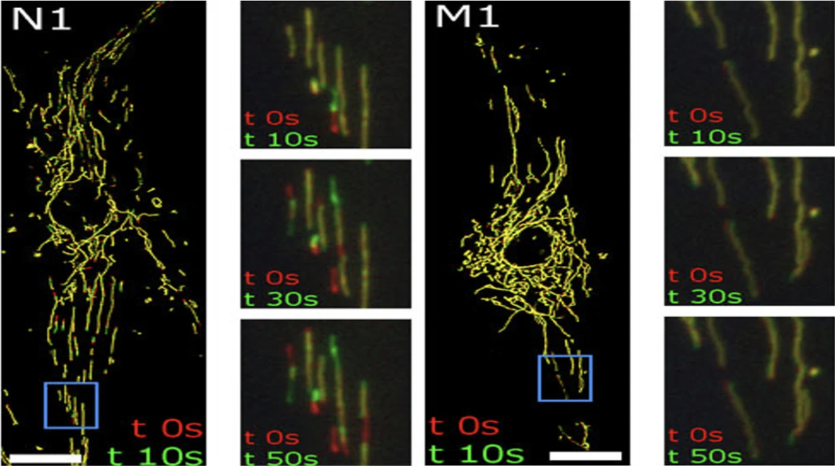
Figure 1. Representative confocal microscopy images of MitoTracker-stained skin fibroblasts demonstrating the strikingly altered mitochondrial distribution in MERRF patient-derived cells compared with controls. Live-cell imaging further reveals markedly reduced mitochondrial motility in MERRF skin fibroblasts, highlighting impaired mitochondrial dynamics associated with the disease phenotype.
Expanding on both this metabolic transition and our recent demonstration that mitochondrial impairment is coupled with synaptic defects in MERRF neurons [4], we find that the legacy of this defective metabolic reprogramming induces a significant delay in early neurite formation and maturation during the terminal stage of neuron differentiation.
Although glycolytic acceleration may sustain early survival, mature cortical neurons strictly demand immense localized ATP for the cycling of synaptic vesicles. Consequently, the unresolved mitochondrial fragmentation and bioenergetic deficit directly translate into impaired Ca2+ buffering in the cytosol, compromised synaptogenesis and defective neurotransmission, which can serve as a precise in vitro manifestation of the neurological disorders often observed in patients with MERRF syndrome.
Precision modeling of mtDNA heteroplasmy thresholds
Mitochondrial diseases are uniquely characterized by mtDNA heteroplasmy, the co-existence of wild-type and mutated mtDNA within a single cell. The management of mitochondrial diseases is exceptionally difficult due to the multi-copy nature of the mitochondrial genome. However, recent advancements have shifted the perspective of mitochondrial medicine, demonstrating that complete clearance of mutant mtDNA is not an absolute prerequisite for making a proper therapeutic choice. Instead, therapeutically manipulating mtDNA heteroplasmy to shift the mutant load just below a specific threshold can fundamentally restore OXPHOS capacity and prevent the ROS overproduction. A pivotal challenge in mitochondrial medicine has been to determine whether heteroplasmy shifting is viable in post-mitotic, highly energy-dependent tissues like the brain. Recent breakthrough studies in mitochondrial disease utilizing the culture cells and in vivo mouse models have successfully addressed this issue using mitochondria-targeted nucleases, specifically mitoTALENs [14].
Engineered to selectively recognize and cleave mutated mtDNA (e.g., m.8344A>G), these nucleases induce the rapid degradation of the pathogenic mitochondrial genome [15]. A recent study demonstrated that AAV-mediated delivery of mitoTALENs can effectively reduce the mtDNA mutation (m.5024C>T) load directly in mammalian neurons in vivo [16]. Because these post-mitotic cells lack robust double-strand break repair mechanisms for the mutant mtDNAs, the cleaved mutant mitochondrial genomes are degraded and the remaining wild-type mtDNAs are allowed to repopulate the mitochondrial network.
This targeted heteroplasmy shift in murine neurons successfully rescued respiratory chain defects, suppressed the ROS overproduction and restored the bioenergetic capacity required for synaptic transmission.
Building on this pivotal proof of concept, our patient-specific MERRF iPSC-derived neurons serve as an ideal preclinical platform to translate this precision gene-editing technology into clinical applications.
Clinical paradigms: from personalized diagnostics to breakthrough therapeutics
The integration of these patient iPSC-derived neurons into clinical practice establishes a powerful platform to expedite the development of personalized medicine and regenerative cell therapies.
3D organoid platforms for functional validation
While non-invasive 2D iPSC cultures provide an invaluable and highly scalable platform for dissecting cell-autonomous mechanisms and validating genetic variants, they can be further complemented to capture the broader, multicellular architecture of the human brain.
Building upon recent successes in modeling other mitochondrial encephalomyopathies, 3D iPSC-derived brain organoids are poised to extrapolate these 2D findings into a complex three-dimensional microenvironment, serving as an advanced preclinical platform for the disease modeling of MERRF syndrome. As highlighted in a comprehensive model of Leigh syndrome [17], 3D organoids bridge the evolutionary gap between lower organism models and human pathology.
Furthermore, recent studies of modeling MELAS (mitochondrial encephalopathy, lactic acidosis, and stroke-like episodes) syndrome emphasize that brain organoids are uniquely equipped to recapitulate the complex multicellular metabolic interplay [18]. At the molecular level, these 3D organoid models have revealed that mtDNA mutations trigger hyperactive Notch signaling, which fundamentally alters neural progenitor cell proliferation and disrupts complex cytoarchitecture, giving rise to the pathological features that remain entirely invisible in standard 2D cultures.
A recent study demonstrated that targeted metabolic intervention, supplementation with the NAD+ precursor nicotinamide riboside (NR), successfully rescued morphological defects of mitochondria and prevented large-scale neuronal loss within the organoid tissue matrix [19]. By integrating targeted interventions (e.g., mitoTALENs or metabolic modulators) into MERRF 3D organoids, researchers can definitively demonstrate how metabolic rescue at the single-neuron level translates into the restoration of the complex cytoarchitecture and network connectivity.
High-throughput screening and the therapeutic repurposing of sildenafil
The urgent need for effective treatments for mitochondrial diseases has driven the adoption of automated high-throughput screening. A recent large-scale iPSC-based screening revealed a novel therapeutic application for sildenafil [20]. Beyond its classical role as a phosphodiesterase type 5 (PDE5) inhibitor, sildenafil significantly alleviates mitochondrial disease phenotypes by attenuating intracellular oxidative stress, boosting mitochondrial biogenesis, and enhancing energy generation. Transitioning this repurposed drug into MERRF-derived neural cell models offers a highly promising strategy to restore redox homeostasis and safeguard synaptic function and structural integrity.
Bioengineered mitochondrial transplantation to overcome translational hurdle via precision organelle tuning
For patients with advanced stages of MERRF syndrome suffering from severe bioenergetic exhaustion, –regenerative medicine offers mitochondrial transplantation. The fundamental premise of mitochondrial transplantation is to physically replace the defective, mutant mtDNA-bearing organelles with healthy exogenous mitochondria to directly reboot cellular bioenergetics. The major bottleneck in translating mitochondrial transplantation into clinical practice has been the difficulty of isolating large amounts of viable, structurally intact, and highly active mitochondria. However, recent bioengineering advancements have introduced organelle-tuning conditions, allowing researchers to fabricate massive yields of functional and energetic mitochondria from donor stem cells [21]. Researchers can use a pre-conditioning environment to maximize the respiratory capacity, optimize membrane potential, and preserve the structural integrity of mitochondria prior to isolation.
The highly active mitochondria increased cellular ATP level and mitochondrial membrane potential in the chondrocytes of osteoarthritis patients. Through advanced bioencapsulation techniques, exogenous mitochondria can be delivered to prevent extracellular degradation, allowing them to be actively internalized by energy-depleted host neurons, and this transplantation strategy has been shown to effectively alleviate functional deficits in both mitochondrial and Parkinson’s disease models [22].
Applying this novel bioprocessing technology to MERRF therapy is a highly promising approach to facilitate efficient cellular uptake of isolated mitochondria by energy-depleted host neurons and achieve the restoration of neural function. Once integrated, they hold the potential to reboot cellular bioenergetics, alleviate OXPHOS defects, and facilitate the electrophysiological recovery of compromised neural networks.
Concluding remarks
Moving beyond the classical paradigm of simple ATP deficiency in mitochondrial diseases, current researchers have increasingly focused on profound developmental aberrations in patients with MERRF syndrome driven by metabolic reprogramming failure. We demonstrated that dysfunctional mitochondria may be accumulated as a result of the stalled and maladaptive mitophagic flux caused by oxidative stress. This mitochondrial structural and metabolic abnormality ultimately precipitates in a severe bioenergetic crisis, leading to profound deficiency in synaptic energy and network connectivity in mature neurons.
We showed that iNSCs and mature neurons differentiated from the iPSCs of patients with MERRF syndrome can recapitulate the biochemical defects, oxidative stress and clinical pathology of the disease and may serve as a platform for the high-throughput screening of novel drugs [4,11].
Ultimately, the integration of advanced 3D organoid modeling with precision interventions, ranging from mitoTALEN-mediated mitochondrial genome editing and old drug repurposing to mitochondrial transplantation, has provided a transformative blueprint and tangible hope for the mitigation of the devastating neurological disorders of mitochondrial diseases.
Acknowledgement
The work described in this article has been supported by grants from the National Science and Technology Council of Taiwan Government (NSTC 111-2320-B-371-001, 112-2320-B-371-001, NSTC 113-2320-B-371-001), intramural research grants (112-CCH-IRP-120, 113-CCH-IRP-094, 114-CCH-HCR-143) from Changhua Christian Hospital, Taiwan. We gratefully acknowledge Prof. Yi-Chao Hsu at Mackay Medical University for the inspiring and insightful discussions during our previous collaborations.
Conflict of interest
The authors declare no competing financial interests or personal relationships that could have appeared to influence the work reported in this paper.
Corresponding author
Address correspondence to Prof. Yau-Huei Wei (e-mail: 181765@cch.org.tw).

Prof. Yau-Huei Wei, Ph.D. is a renowned mitochondrial biologist and a pioneer in mitochondrial medicine whose research has advanced the understanding of mitochondrial diseases, aging, and mitochondrial genetics. He served as the Founding President of Mackay Medical College and currently directs the Center for Mitochondrial Medicine and Free Radical Research at Changhua Christian Hospital. Professor Wei has published extensively on mitochondrial dysfunction, oxidative stress, and mitophagy, and has played a key role in promoting mitochondrial research across Asia.
References:
- Finsterer J, Zarrouk-Mahjoub S, Shoffner JM. MERRF Classification: Implications for diagnosis and clinical trials. Pediatr Neurol. 2018;80:8–23.
- Chou SJ, Tseng WL, Chen CT, Lai YF, Chien CS, Chang YL, et al. Impaired ROS scavenging system in human induced pluripotent stem cells generated from patients with MERRF syndrome. Sci Rep. 2016;6:23661.
- Chen YC, Tsai CL, Wei YH, Wu YT, Hsu WT, Lin HC. ATOH1/RFX1/RFX3 transcription factors facilitate the differentiation and characterisation of inner ear hair cell-like cells from patient-specific induced pluripotent stem cells harbouring A8344G mutation of mitochondrial DNA. Cell Death Dis. 2018;9:437.
- Wu YT, Tay HY, Yang JT, Liao HH, Ma YS, Wei YH. Mitochondrial impairment and synaptic dysfunction are associated with neurological defects in iPSCs-derived cortical neurons of MERRF patients. J Biomed Sci. 2023;30(1):70.
- Sinenko SA, Tomilin AN. Metabolic control of induced pluripotency. Front Cell Dev Biol. 2024;11:1328522.
- Knobloch M, Jessberger S. Metabolism and neurogenesis. Curr Opin Neurobiol. 2017;42:45–52.
- Landoni JC, Lycas MD, Macuada J, Stepp W, Jaccard R, Obara CJ, et al. Pearling drives mitochondrial DNA nucleoid distribution. Science. 2026; 392(6793):102–9.
- Sturm G, Hake K, Lefebvre AEYT, Rux CJ, Ivanova D, Millett-Sikking A, et al. The biophysical mechanism of mitochondrial pearling. Mol Biol Cell. 2025;36(11): ar142.
- Cartes-Saavedra B, Lagos D, Macuada J, Arancibia D, Burté F, Sjöberg-Herrera MK, et al. OPA1 disease-causing mutants have domain-specific effects on mitochondrial ultrastructure and fusion. Proc Natl Acad Sci USA. 2023;120:e2207471120.
- Zhang L, Trushin S, Christensen TA, Bachmeier BV, Gateno B, Schroeder A, et al. Altered brain energetics induces mitochondrial fission arrest in Alzheimer’s disease. Sci Rep. 2016;6:18725.
- Wu YT, Tay HY, Liao HH, Yang JT, Wei YH. Role of oxidative stress in the susceptibility to mitophagy of the skin fibroblasts and iPSC-derived neurons of patients with MERRF syndrome. Mitochondrion. Published online 19 May 2026:102171. doi: 10.1016/j.mito.2026.102171.
- Chan, DC. Mitochondrial dynamics and its involvement in disease. Annu Rev Pathol. 2020;15:235–59.
- Lorenz C, Lesimple P, Bukowiecki R, Zink A, Inak G, Mlody B, et al. Human iPSC-derived neural progenitors are an effective drug discovery model for neurological mtDNA disorders. Cell Stem Cell. 2017;20(5):659–74.e9.
- Zekonyte U, Bacman SR, Moraes CT. DNA-editing enzymes as potential treatments for heteroplasmic mtDNA diseases. J Intern Med. 2020;287(6):685–97.
- Hashimoto M, Bacman SR, Peralta S, Falk MJ, Chomyn A, Chan DC, et al. MitoTALEN: A general approach to reduce mutant mtDNA loads and restore oxidative phosphorylation function in mitochondrial diseases. Mol Ther. 2015;23(10):1592–9.
- Bacman SR, Barrera-Paez JD, Pinto M, Van Booven D, Stewart JB, Griswold AJ, et al. mitoTALEN reduces the mutant mtDNA load in neurons. Mol Ther Nucleic Acids. 2024;35(1):102132.
- Henke MT, Prigione A, Schuelke M. Disease models of Leigh syndrome: From yeast to organoids. J Inherit Metab Dis. 2024;47(6):1292–321.
- Winanto, Khong ZJ, Soh BS, Fan Y, Ng SY. Organoid cultures of MELAS neural cells reveal hyperactive Notch signaling that impacts neurodevelopment. Cell Death Dis. 2020;11(3):182.
- Hong Y, Zhang Z, Yangzom T, Chen A, Lundberg BC, Fang EF, et al. The NAD+ precursor nicotinamide riboside rescues mitochondrial defects and neuronal loss in iPSC derived cortical organoid of Alpers’ disease. Int J Biol Sci. 2024;20(4):1194–217.
- Zink A, Dai DF, Wittich A, Henke MT, Pedrotti G, Heiduschka S, et al. Pluripotent stem-cell-based screening uncovers sildenafil as a mitochondrial disease therapy. Cell. 2026;189(6):1656–79.e42.
- Chen X, Zhou Y, Yao W, Gao C, Sha Z, Yi J, et al. Organelle-tuning condition robustly fabricates energetic mitochondria for cartilage regeneration. Bone Res. 2025;13(1):37.
- Du S, Long Q, Zhou Y, Fu J, Wu H, Yang L, et al. Transplantation of encapsulated mitochondria alleviates dysfunction in mitochondrial and Parkinson’s disease models. Cell. 2026;189(10):2821–33.e23.

Leave a comment